October is Breast Cancer Awareness Month—a useful reminder to take an active role in your breast health, understand your imaging options, and keep recommended screening up to date.
At Breast Thermography International (BTI), we believe that understanding an examination is just as important as scheduling it. If you have wondered what breast thermography measures, what the colors mean, or what happens during an appointment, this guide explains the basics. You can also explore our breast thermography overview.
What is breast thermography?
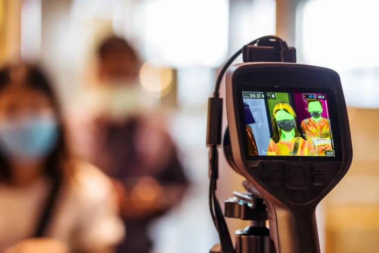
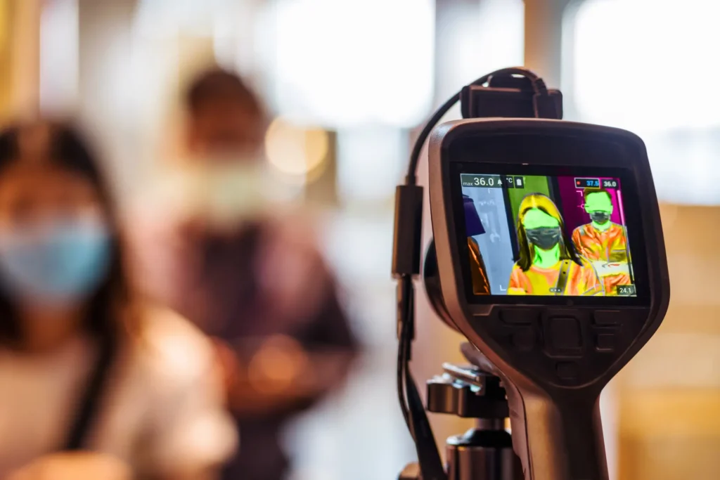
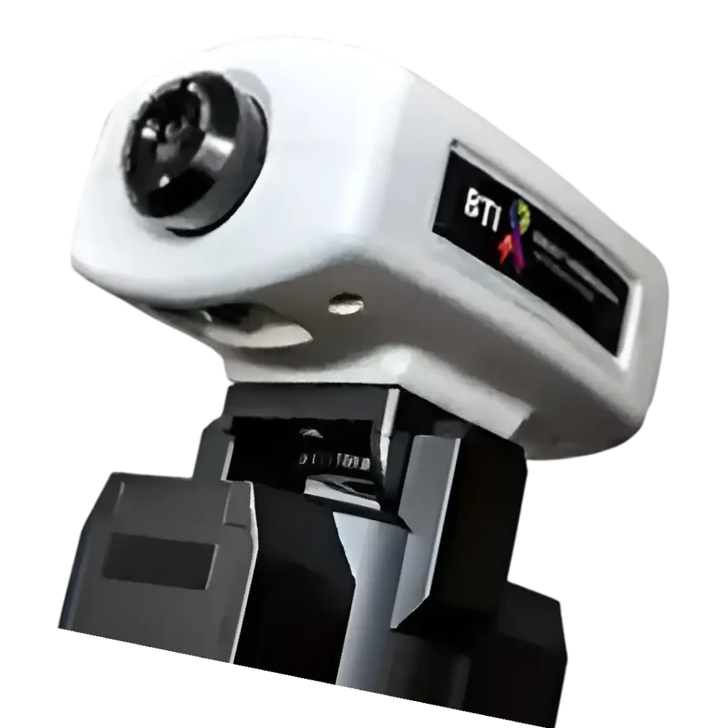
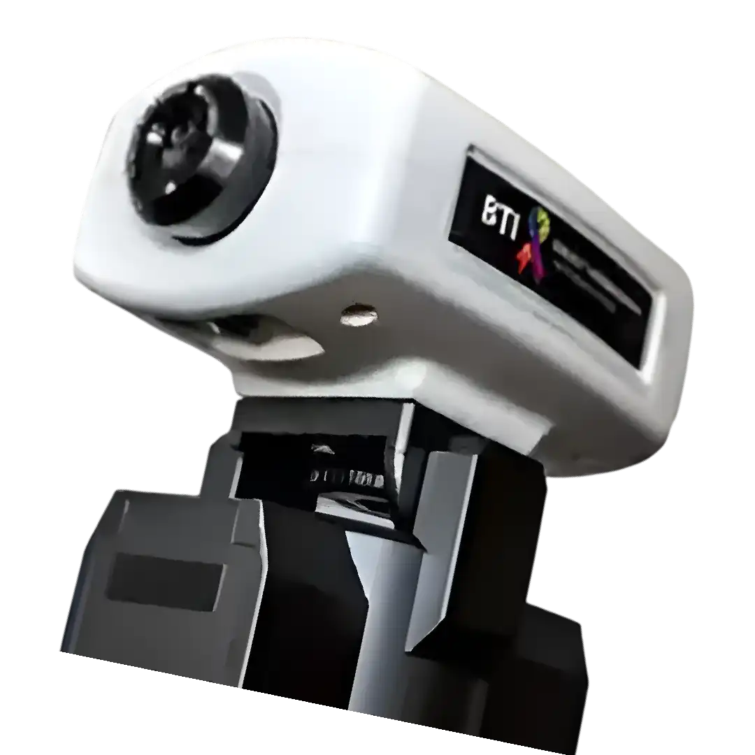
Breast thermography uses a high-resolution thermography camera to detect infrared energy emitted by the skin and create a map of surface-temperature patterns. The camera records heat; it does not send radiation into your body.
The examination is noncontact and does not require breast compression or use ionizing radiation. It measures temperature at the skin surface rather than producing an image of the internal structure of the breast.
How does thermography work?
Skin temperature is influenced by blood flow and other physiological processes. A thermographic examination records these temperature patterns under controlled conditions so an interpreting physician can assess the images in the context of your symptoms and medical history.
The colors on a thermal image represent temperatures within the selected display scale. A warmer color does not automatically mean cancer, and a cooler color does not establish that tissue is healthy. The color palette is a way to display measurements—not a diagnosis.
Temperature differences can have several explanations, including normal variation, hormonal influences, inflammation, and environmental or preparation factors. Thermography alone cannot determine the cause of a finding.
What happens during an appointment?
- Review your history. Share breast symptoms, previous procedures, relevant medical history, and earlier imaging or thermography reports.
- Allow time to acclimate. You will spend time in a temperature-controlled room before imaging. This helps reduce the influence of recent clothing, activity, and outside temperatures.
- Capture the images. The technician records a series of views with the infrared camera without touching or compressing the breasts.
- Review the report. The images are submitted for interpretation. Discuss findings and any recommended next steps with your healthcare professional.
Read what to expect at your thermography visit and follow the specific preparation instructions supplied by your imaging location. Exercise, heat exposure, and products applied to the skin can affect temperature measurements. Do not stop prescribed medication unless your treating clinician directs you to do so.
Why might previous thermal images be compared?
When earlier studies are available, the interpreting physician may compare surface-temperature patterns over time. Consistent preparation and imaging conditions help make those comparisons more meaningful.
Comparison can document whether recorded patterns appear stable or have changed. However, stable or normal thermographic findings do not rule out breast cancer and should not be used to postpone recommended screening or evaluation of a new symptom.
Where does thermography fit into breast care?
Different examinations measure different things. Mammography uses low-dose X-rays to examine breast structure and is the standard screening test for most women. Ultrasound or MRI may be recommended for particular symptoms, imaging findings, or risk factors.
Thermography cannot diagnose or rule out breast cancer. It is not a substitute for mammography or other clinically indicated tests. The FDA states that thermography devices have been cleared only for adjunctive use alongside a primary test such as mammography. This clearance does not establish thermography as an effective standalone breast cancer screening method.
If you choose thermography, discuss its limitations with your healthcare professional and keep your recommended screening plan current. Your age, personal history, family history, and other risk factors should guide that plan.
Breast changes that deserve medical attention
Become familiar with how your breasts usually look and feel. Contact a healthcare professional if you notice:
- A new lump or area of thickening in the breast or underarm.
- A change in breast size or shape.
- Skin dimpling, persistent redness, or other new skin changes.
- A newly inverted nipple or unexpected nipple discharge.
- New, persistent pain in one area.
Many breast changes have benign causes, but they still deserve appropriate evaluation. Do not wait for a thermography appointment or rely on a previous normal result to assess a new concern.
Frequently asked questions
Does breast thermography hurt?
The imaging itself is noncontact and requires no compression. You will need to uncover the area being imaged and hold the positions requested by the technician.
Does it use radiation?
It does not use ionizing radiation. The camera detects infrared energy naturally emitted by your skin.
Can it replace my mammogram?
No. Keep mammography and any other imaging recommended by your healthcare professional up to date.
What should I do after receiving my report?
Discuss it with your healthcare professional, including any follow-up recommendations. A thermal finding may require clinical assessment or other testing; a normal report does not exclude cancer.
Make breast awareness a year-round habit
Use October as a reminder to review your family history, speak up about changes, and make an informed breast-care plan with your clinician. Awareness is most useful when it leads to appropriate action.
To learn about an appointment through the BTI network, find a BTI thermography location and contact a participating technician for preparation instructions and scheduling.
This article provides general education and does not replace individual medical advice.