- Home
- Thermography Case Studies
Thermography Case Studies
9x7x5 cm Tumor
Thermal Imaging Analysis of Tumor Activity
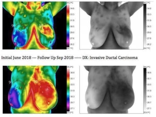
6 Month Follow-up
Thermographic Evidence of Aggressive Breast Cancer Development
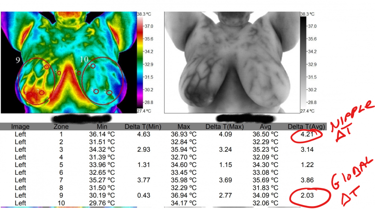
Lymphatic Involvement
Infrared Indicators of Lymphatic Cancer Progression
- Detects abnormal heat patterns in breast and abdomen.
- Indicates possible lymphatic system involvement.
- Tracks spread of cancer through thermal imaging.
- Highlights inflammation linked to cancer cells.
- Supports diagnosis and staging of breast cancer.
- Identifies potential for metastasis early.
- Visualizes cancer progression in real time.
- Non-invasive tool for monitoring cancer changes.
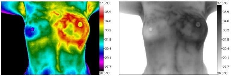
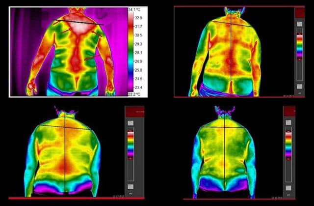
Thermal Effects of Aberrant Posture
Detecting Inflammation from Poor Posture Using Thermal Imaging
Thermography can be used for more than just breast studies. This full body thermography case study shows the stress of poor posture on the human body. Here, a lateral thoracic translation can be seen. The red areas are areas of hyperthermia (increased heat), indicating biomechanical stress in the form of inflammation. Full body thermography is an effective way to assess for and monitor improvement of musculoskeletal dysfunction.
9x7x5 cm Tumor
Full Body Thermography to Monitor Treatment Outcomes
Thermography can effectively monitor progress in neuromuscular conditions. This full body thermography case study shows the improvements made by a Chiropractic patient. The physician treated her neuromuscular complaints and took follow up scans at 12 visit (1 month) increments. Thermography is a valuable tool for assessing and monitoring improvement in neuromuscular conditions.
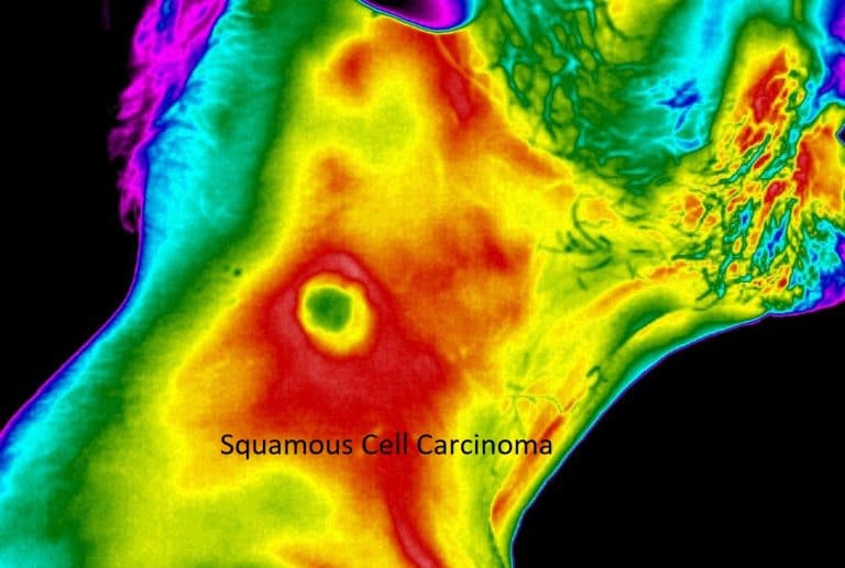
Squamous Cell Carcinoma
Thermographic Detection of Squamous Cell Carcinoma
This thermography case study shows a squamous cell carcinoma that was caused by radiation therapy from a contralateral lymphoma. Notice the areas of heat surrounding the center. On thermography, areas of excessive heat (red and white) could be indicative of angiogenesis. Angiogenesis is the formation of new blood vessels which results in increased blood flow to the area, supplying tumors and cancer cells with the nutrients they need for growth and reproduction.
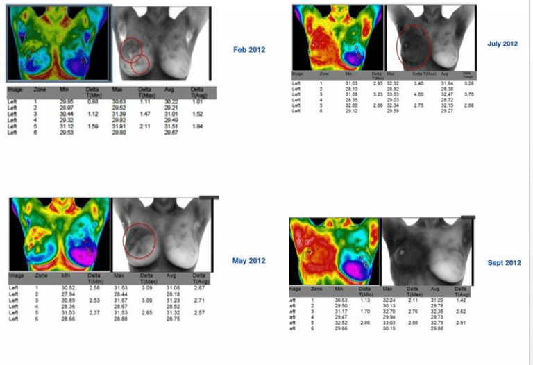
Inflammatory Breast Cancer
Radiation-Free Imaging for Aggressive Breast Cancer Cases
This breast thermography case study shows the rapid progression that inflammatory breast cancer can present with. This patient attempted several alternative treatments. She refused surgery and traditional forms of cancer treatment, against the recommendations of her doctor. Breast thermography can aid in monitoring breast cancer throughout the treatment process. Thermography is 100% safe and radiation free which allows for imaging as frequently as necessary for peace of mind while undergoing traditional forms of cancer treatment.